Volume 14, Issue 2 (Spring 2026)
Iran J Health Sci 2026, 14(2): 183-192 |
Back to browse issues page



Ethics code: 1401.294
Clinical trials code: 0


Download citation:
BibTeX | RIS | EndNote | Medlars | ProCite | Reference Manager | RefWorks
Send citation to:


BibTeX | RIS | EndNote | Medlars | ProCite | Reference Manager | RefWorks
Send citation to:
Fattahian A, Amid N, Ghaemian A, Ghadirzadeh E, Moosazadeh M. Comparative Risk Factors of Premature Versus Late-Onset Coronary Artery Disease: A Case-control Study. Iran J Health Sci 2026; 14 (2) :183-192
URL: http://jhs.mazums.ac.ir/article-1-1218-en.html
URL: http://jhs.mazums.ac.ir/article-1-1218-en.html


Gastrointestinal Cancer Research Center, Non-communicable Disease Institute, Mazandaran University of Medical Sciences, Sari, Iran. , mmoosazadeh1351@gmail.com
Full-Text [PDF 777 kb]
(225 Downloads)
| Abstract (HTML) (998 Views)
Full-Text: (86 Views)
Introduction
Cardiovascular disease (CVD) is among the leading causes of mortality worldwide [1]. According to the World Health Organization (WHO), approximately 17.9 million people die annually from CVDs, accounting for nearly 32% of all global deaths [1]. In Iran, CVD is the leading cause of death and represents the most prevalent group of chronic diseases, with an estimated prevalence of 21% [2]. Among CVDs, coronary artery disease (CAD) is one of the major contributors to morbidity and mortality worldwide [3]. CAD is characterized by reduced blood and oxygen supply to the myocardium due to obstruction of the coronary arteries, resulting in an imbalance between myocardial oxygen supply and demand.
Despite advances in prevention and treatment, CAD is expected to remain a leading cause of disability and premature death worldwide through 2030 [4]. Recent evidence suggests that the age of CAD onset has decreased significantly, particularly in Asian countries [5]. Consequently, the term premature CAD (PCAD) has emerged to describe CAD occurring at a younger age. PCAD is commonly defined as acute myocardial infarction or symptomatic myocardial ischemia with coronary stenosis ≥70% occurring in men younger than 45 years and women younger than 55 years [6, 7]. In contrast, its usual late-onset type CAD (LCAD) generally occurs in men older than 55 years and women older than 65 years [6]. Previous studies have reported that the prevalence of CAD is approximately 37% among younger men (<55 years) and women (<65 years), compared with 67% among older individuals [8].
Ischemic heart disease in younger populations has become an important cause of morbidity and mortality, particularly in developing countries, and recent studies have reported increasing rates of PCAD and myocardial infarction in these populations [5]. In contrast, the incidence of myocardial infarction among middle-aged and older patients (contributing to LCAD) has declined in recent years [9]. One possible explanation for this trend may be differences in the risk factor profiles of PCAD and LCAD [9, 10].
Several studies have identified significant associations between LCAD and risk factors such as cigarette smoking, family history of CAD, substance abuse, dyslipidemia (DLP), hypertension (HTN), obesity, sedentary lifestyle, and diabetes mellitus (DM) [11]. However, relatively few studies have specifically investigated the risk factors associated with PCAD. A large systematic study comparing patients with PCAD to individuals with normal coronary arteries reported that DLP (52%), cigarette smoking (66%), and family history of CAD (90%) were among the most important risk factors associated with PCAD [12]. Other studies have similarly identified smoking, elevated cholesterol levels, increased low-density lipoprotein (LDL), HTN, hypertriglyceridemia, metabolic syndrome, and social determinants as major contributors to PCAD [13-15]. Nevertheless, due to the limited number of available studies, the risk factor profile of PCAD remains insufficiently understood.
Since PCAD is a multifactorial disease, further research comparing the risk factors of PCAD and LCAD is warranted [8]. A review of the existing literature indicates that most previous studies have compared patients with PCAD to those without CAD, while few have directly compared PCAD with LCAD. Understanding potential differences in risk factor profiles between these two groups is essential for improving prevention strategies and reducing CAD-related morbidity and mortality at younger ages. Therefore, the present study aimed to compare the risk factors associated with premature and late CAD among patients referred to Fatemeh-Zahra Medical Center (Mazandaran Heart Center).
Materials and Methods
Study design and participants
This case–control study was conducted among patients diagnosed with PCAD and LCAD who were admitted to the Emergency Department of Fatemeh-Zahra Heart Center during 2021-2022. The diagnosis of CAD was confirmed based on coronary angiography findings. The PCAD group included men younger than 45 years and women younger than 55 years, whereas the LCAD group consisted of men older than 45 years and women older than 55 years. Patients with incomplete clinical data or a history of chronic hepatitis, severe renal artery stenosis, severe infections, connective tissue diseases, malignancy, hematologic disorders, pregnancy or breastfeeding, long-term use of contraceptive medications, secondary HTN, or endocrine disorders, such as thyroid dysfunction or adrenal cortical disease, were excluded from the study.
Sampling
To determine the sample size, the results of the study by Kazemi et al [16] were used. In the referenced study, the prevalence of HTN in the case and control groups was reported as 32.98% (approximately 33%) and 13.83% (approximately 14%), respectively. Considering these values, a 95% confidence level, 90% statistical power, and a two-sided test, and using the formula for comparing two proportions along with G*Power (version 3.1.9.2), the minimum required sample size was estimated to be 206 participants (103 individuals in each group).
Participants were recruited using convenience sampling. After obtaining approval from the Deputy of Research at Mazandaran University of Medical Sciences, eligible patients presenting with chest pain and suspected CAD were assessed for study inclusion and exclusion criteria. Patients who met the inclusion criteria and had angiography-confirmed CAD were categorized into either the PCAD or LCAD group and subsequently compared.
Coronary angiography and CAD definition
Coronary angiography was performed using the standard Judkins technique in seven conventional views [17]. For the left coronary artery, angiographic imaging was obtained in the left anterior oblique cranial, left anterior oblique caudal, right anterior oblique cranial, and right anterior oblique caudal positions. For the right coronary artery, imaging was performed in the left anterior oblique, right anterior oblique, and cranial positions. Two experienced cardiologists independently evaluated all angiographic findings. CAD was defined as stenosis >50% in the main coronary artery or its major branches. The degree of stenosis in the left main (LM) coronary artery, left anterior descending artery, circumflex artery, and right coronary artery was assessed. Major branches, including the diagonal and obtuse marginal branches, were also evaluated as part of major coronary artery involvement.
Data collection and measurements
Data were collected using a researcher-designed checklist consisting of demographic, clinical, anthropometric, and laboratory variables. Demographic information included age, sex, educational status, socioeconomic status, and residence. The clinical data included underlying diseases, history of anxiety or depression, major life events, medication history, family history of CAD, smoking status, alcohol consumption, and substance abuse.
Following the completion of medical history taking and physical examination, anthropometric measurements were obtained from all participants. Height, weight, waist circumference (WC), and hip circumference (HC) were measured using standard methods. WC was measured at the narrowest circumference between the iliac crest and the lower rib margin at the end of normal expiration, while HC was measured at the maximum prominence of the buttocks. The waist-to-hip ratio (WHR) was subsequently calculated. Body mass index (BMI) was calculated as weight in kilograms divided by the square of height in meters (kg/m²). Blood pressure (BP) was measured in the right arm after at least 5 minutes of rest. Two measurements were obtained at intervals of at least 1 minute, and the average value was recorded for analysis.
Peripheral venous blood samples were collected after 10 hours of overnight fasting without water restriction. Routine biochemical analyses, including fasting blood sugar (FBS), lipid profile, plasma creatinine, liver enzymes, C-reactive protein (CRP), vitamin D levels, and viral hepatitis markers, were measured using an automated analyzer with standard laboratory methods.
Statistical analysis
Data analysis was performed using IBM SPSS Statistics version 21 (IBM Corporation, USA). Data were presented as Mean±SD for continuous variables and frequency (percentage) for categorical variables. The normality of data distribution was assessed using the Kolmogorov–Smirnov test. Continuous variables with normal distribution were compared using the independent-samples t-test, whereas non-normally distributed variables were analyzed using the Mann–Whitney U test. Categorical variables were compared using the chi-square (χ²) test. Multivariate logistic regression analysis was performed to identify risk factors associated with PCAD and LCAD, and adjusted odds ratios (ORs) with 95% confidence intervals (CIs) were calculated. A P<0.05 was considered statistically significant.
Results
The demographic and clinical characteristics of the study participants are presented in Table 1.
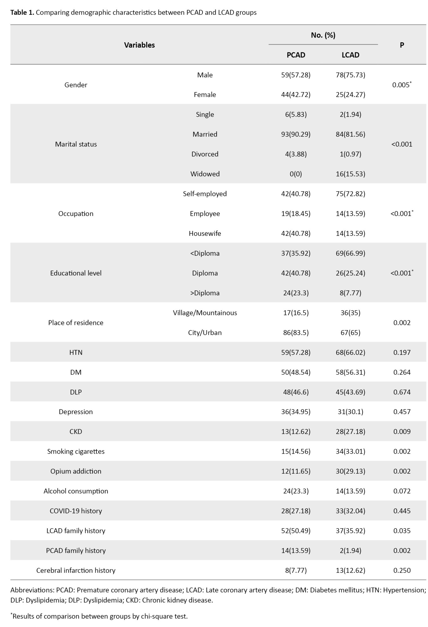
Male participants constituted the majority of both the PCAD and LCAD groups; however, the proportion of men was significantly higher in the LCAD group than in the PCAD group (75.73% vs 57.28%, P=0.005). Most participants in both groups were married, although widowed status was more common among patients with LCAD (P<0.001).
Significant differences were observed between the two groups regarding occupation, educational level, place of residence, chronic kidney disease (CKD), cigarette smoking, opium addiction, and family history of CAD (P<0.05 for all). Patients in the PCAD group were more likely to have higher educational attainment and a positive family history of both LCAD and PCAD, whereas patients in the LCAD group more frequently had lower educational levels, rural residence, CKD, cigarette smoking, and opium addiction.
No significant differences were found between the groups with respect to HTN, DM, DLP, depression, alcohol consumption, COVID-19 history, or previous cerebral infarction (P>0.05).
Regarding coronary artery involvement, single-vessel disease (SVD) was more common among patients with PCAD, whereas three-vessel disease (3VD) and LM involvement were more frequent in the LCAD group.
Comparisons of anthropometric and laboratory parameters between the two groups are summarized in Table 2.
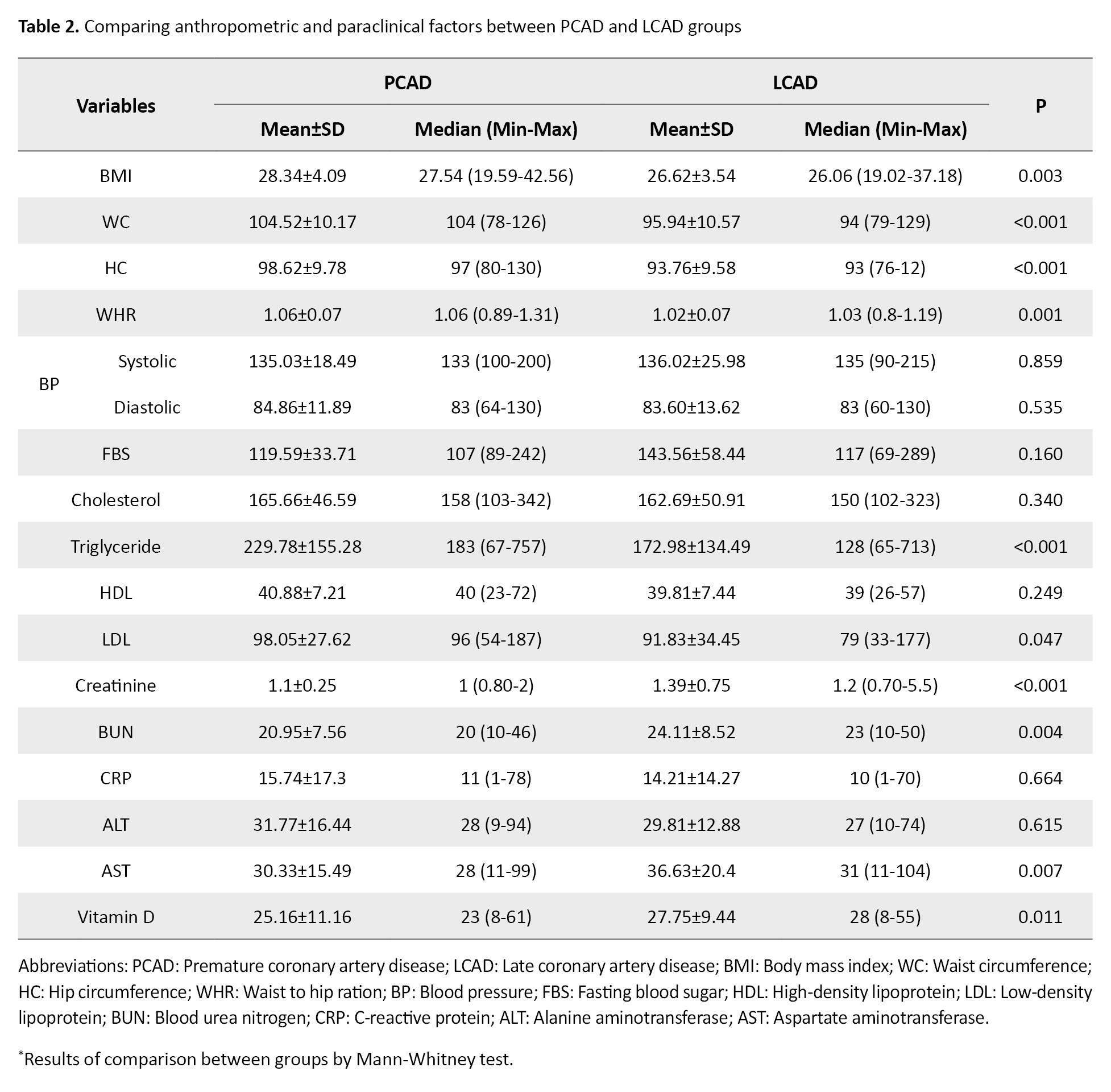
Patients with PCAD had significantly higher BMI, WC, HC, WHR, triglyceride, and LDL levels, compared with patients with LCAD (P<0.05). In contrast, serum creatinine, blood urea nitrogen (BUN), aspartate aminotransferase (AST), and vitamin D levels were significantly higher in the LCAD group than in the PCAD group (P<0.05). No statistically significant differences were observed between the groups regarding systolic and diastolic BP, FBS, total cholesterol, HDL, CRP, or alanine aminotransferase (ALT) levels (P<0.05).
Multivariable logistic regression analysis was performed to identify factors independently associated with PCAD compared with LCAD (Table 3).
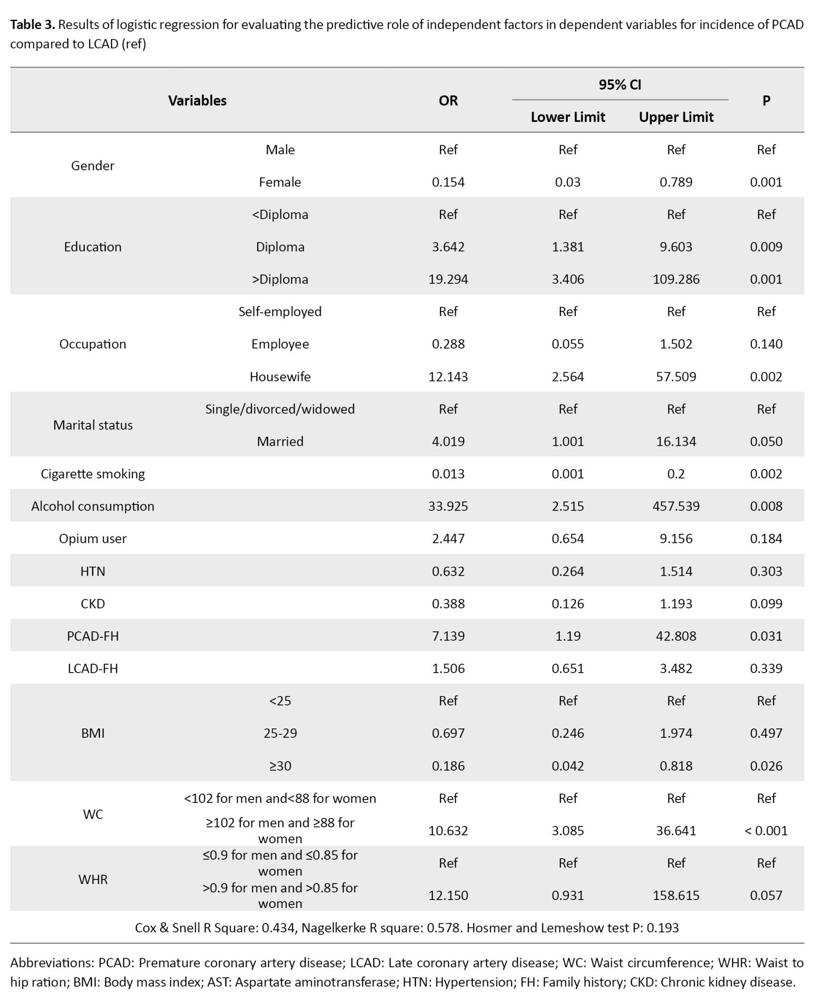
Higher educational level was significantly associated with increased odds of PCAD. Compared with participants with education below diploma level, individuals with diploma-level education had 3.64-fold higher odds of PCAD (OR=3.642, 95% CI, 1.381%, 9.603%, P=0.009), while those with education above diploma level had 19.29-fold higher odds (OR=19.294, 95% CI, 3.406%, 109.286%, P=0.001).
Housewife status (OR=12.143, 95% CI, 2.564%, 57.509%, P=0.002), alcohol consumption (OR=33.925, 95% CI, 2.515%, 457.539%, P=0.008), family history of PCAD (OR=7.139, 95% CI, 1.19%, 42.808%, P=0.031), and elevated WC (OR=10.632, 95% CI, 3.085%, 36.641%, P<0.001) were also independently associated with a higher risk of PCAD.
Female sex was associated with lower odds of PCAD than male sex (OR=0.154, 95% CI, 0.03%, 0.789%, P=0.001). Cigarette smoking was inversely associated with PCAD and, therefore, more strongly associated with LCAD (OR=0.013 [95% CI, 0.001%, 0.2%], P=0.002). Similarly, obesity (BMI ≥30 kg/m²) was associated with lower odds of PCAD (OR=0.186 [95% CI, 0.042%, 0.818%], P=0.026).
Although a borderline association was observed between higher WHR and PCAD, the relationship did not reach statistical significance (OR=12.150 [95% CI: 0.931–158.615], P=0.057). The final regression model demonstrated acceptable goodness-of-fit according to the Hosmer–Lemeshow test (P=0.193), with Cox and Snell R² and Nagelkerke R² values of 0.434 and 0.578, respectively.
Discussion
The present study aimed to compare the demographic, clinical, anthropometric, and laboratory risk factors associated with PCAD and LCAD among patients with angiography-confirmed CAD. The findings demonstrated significant differences between the two groups in several risk factors. Patients with PCAD had significantly higher BMI, WC, HC, triglyceride and LDL levels, and WHR, whereas creatinine, BUN, AST, and vitamin D levels were higher among patients with LCAD. In addition, multivariable logistic regression analysis showed that higher educational level, housewife status, alcohol consumption, family history of PCAD, and elevated WC were more strongly associated with increased odds of PCAD (rather than LCAD), while cigarette smoking was more strongly associated with LCAD. These findings suggest that PCAD and LCAD may have distinct risk factor profiles that should be considered in preventive and therapeutic strategies.
The results of the study by Al-Khlaiwi et al. are consistent with the results of the present study, showing that family history of CAD was common among patients with PCAD diagnosis, with a pooled estimated prevalence of 19% [18], which indicates the strong influence of family history of CAD on the premature occurrence of CAD.
The findings of the present study were partly consistent with the results reported by Khoja et al. [19]. Similar to their study, we found that patients with PCAD had significantly higher BMI, triglyceride, and LDL levels, as well as central obesity indices, including WC and WHR, compared to patients with LCAD. In addition, both studies demonstrated that a positive family history of CAD was more common among patients with premature disease, supporting the important role of genetic predisposition in the early onset of CAD. These similarities suggest that metabolic abnormalities and hereditary factors may contribute substantially to the development of CAD at younger ages.
However, several differences were observed between the two studies. Khoja et al. reported that smoking was more prevalent among patients with early-onset coronary heart disease, whereas in the present study cigarette smoking and opium addiction were significantly more common among patients with LCAD. In addition, Khoja et al. found lower systolic BP and lower prevalence of HTN and DM among younger patients, while our study showed no significant differences between the PCAD and LCAD groups in BP, HTN, or diabetes. Furthermore, Khoja et al. reported lower HDL levels among younger patients, whereas no significant difference in HDL levels was observed in our population.
Several factors may explain these discrepancies. First, differences in study populations and sociocultural characteristics may have influenced the findings. Our study was conducted in an Iranian population in which smoking and opium use are more prevalent among older individuals, potentially explaining the stronger association between smoking and LCAD. Second, variations in dietary habits, physical activity, healthcare access, and cardiovascular risk management among populations may account for differences in HTN, diabetes, and lipid profiles. Third, differences in the definitions of premature disease, inclusion criteria, and study design could also contribute to inconsistent results. For example, Khoja et al. included individuals with early onset coronary heart disease under 65 years of age, whereas the present study used stricter age criteria to define PCAD (<45 years for men and <55 years for women). Finally, the relatively small sample size of the present study compared with the pooled meta-analytic data used by Khoja et al. may have limited the statistical power to detect some associations.
The findings of the present study were consistent with those reported by Mahjoob et al. [20], particularly regarding the distinct clinical and angiographic profiles of younger and older patients with CAD. Both studies demonstrated that younger patients with CAD were more likely to have a positive family history of CAD. In contrast, older patients had a higher prevalence of chronic comorbidities such as HTN, DM, and more extensive coronary involvement.
In terms of angiographic findings, our results were consistent with those of Mahjoob et al. Both studies found that SVD was more prevalent among younger patients, whereas multi-vessel disease, particularly 3VD, was more common among older patients. Mahjoob et al. reported significantly higher rates of two-vessel disease, 3VD, and LAD and RCA involvement among older individuals, while younger patients more commonly exhibited SVD. Similarly, in the present study, SVD was more frequently observed in the PCAD group, whereas 3VD and LM involvement were more common among patients with LCAD. These findings suggest that CAD in younger individuals is often less extensive and may represent the earlier stages of atherosclerotic disease progression.
Regarding metabolic and anthropometric factors, some similarities and differences were identified. In our study, BMI, WC, triglyceride levels, LDL levels, and WHR were significantly higher among patients with PCAD, indicating a stronger contribution of obesity-related and metabolic risk factors in younger patients. However, Mahjoob et al. did not observe significant differences in LDL or triglyceride abnormalities between younger and older groups. Our study evaluated continuous lipid and anthropometric variables, whereas Mahjoob et al. categorized lipid abnormalities using predefined cut-off values, potentially reducing sensitivity to detect intergroup differences. Our study also included sex-specific definitions for PCAD and LCAD, which may have influenced the distribution of metabolic and cardiovascular risk factors between groups.
Our study showed that males were significantly more prevalent in the LCAD group, while female sex was associated with a protective effect for PCAD in regression analysis. Jamil et al. [21] also demonstrated a strong male predominance among patients with atherosclerotic CAD. Both studies reinforce the known protective role of female sex hormones before menopause. Estrogen improves endothelial function, lipid metabolism, and vascular compliance, thereby delaying the onset of clinically significant CAD in women. The results of Jamil et al.’s study also showed that the risk factors for PCAD included diabetes with an OR of 1.98, hyperlipidemia with an OR of 1.85, and history of smoking with an OR of 2.93. However, the findings of Jamil et al. are not directly comparable to our results because their control group consisted of young ACS patients with angiographically normal coronary arteries, whereas our comparator group consisted of older patients with established CAD (LCAD). Consequently, their study primarily evaluated determinants of CAD presence, while our study evaluated determinants associated with earlier CAD onset.
The results of the study by Zhang et al. (2018) revealed high levels of cholesterol, LDL, and HDL as risk factors for late CAD, which was similar to the present study [22]; therefore, it could be said that lipid profiles are effective and preventable factors in the occurrence of PCAD.
One of the inconsistencies among previous studies is the variation in the age criteria used to classify patients into premature and late-onset CAD groups, which may influence the identification and reporting of risk factors associated with these groups. Based on the findings of different studies, it is essential that future investigations evaluating factors associated with CAD and PCAD adopt standardized age ranges to enable more accurate comparisons and improve the reliability and consistency of reported results.
Overall, based on findings from various studies and their comparison with previous research, it appears that the identification of all risk factors associated with LCAD and PCAD remains incomplete. One possible explanation for this issue is the limited number of studies directly comparing risk factors between LCAD and PCAD patients. According to the literature reviewed by the researchers, most previous studies have compared patients with premature or late CAD to healthy or non-CAD populations rather than directly comparing the two CAD groups. Consequently, there is still a considerable gap in knowledge regarding the differential risk factor profiles of LCAD and PCAD patients. In the present study, several sociodemographic variables, including occupation, educational level, and marital status, were identified as factors associated with LCAD and PCAD. However, these variables have not been consistently reported in previous studies. This discrepancy may be attributable to differences in data collection tools, study designs, and the quality or completeness of patients’ medical records, which may have resulted in some relevant variables being overlooked or insufficiently evaluated. Therefore, future studies should consider incorporating comprehensive, standardized data-collection methods to better evaluate the role of sociodemographic factors in the development of premature and late-onset CAD.
Conclusion
In conclusion, PCAD and LCAD demonstrate distinct demographic, metabolic, and clinical risk profiles. Central obesity, DLP, and family history were more strongly associated with PCAD, whereas smoking and renal dysfunction were predominant in LCAD. Recognition of these differences may support prevention, earlier diagnosis, and individualized management strategies for CAD.
Limitations
Since the present study was conducted on a relatively small sample size and within a single medical center, the findings may not be generalizable to all patients with PCAD and LCAD. Therefore, future multicenter studies with larger populations are recommended to improve the generalizability and external validity of these findings. Furthermore, similar to many observational studies, the potential effects of confounding or unmeasured variables cannot be completely excluded. Given the limited number of studies evaluating risk factors associated with premature and late CAD, as well as the inconsistent findings reported in the literature, further research is warranted. Future studies involving larger patient cohorts presenting to medical centers for CAD evaluation are recommended to more accurately identify and compare the risk factors associated with PCAD and LCAD, and to determine whether the shared risk factors observed between the two groups represent true associations rather than incidental findings. Convenience sampling may have introduced selection bias, limiting representativeness and reducing the generalizability of findings to broader CAD populations.
Ethical Considerations
Compliance with ethical guidelines
This study was approved by the Research Ethics Committee of Mazandaran University of Medical Sciences, Sari, Iran (Code: IR.MAZUMS.REC.1401.294). Written informed consent was obtained from all participants.
Funding
This study was conducted with partial financial support from the Mazandaran University of Medical Sciences, Sari, Iran (Grant No.: 9231).
Authors contributions
Conceptualization: Alireza Fattahian and Nakisa Amid; Data curation: Ali Ghaemian and Nakisa Amid; Formal analysis: Mahmood Moosazadeh and Erfan Ghadirzadeh; Methodology: Alireza Fattahian and Mahmood Moosazadeh; Project administration: Nakisa Amid and Ali Ghaemian; Resources: Ali Ghaemian, Alireza Fattahian, and Mahmood Moosazadeh; Software: Erfan Ghadirzadeh and Mahmood Moosazadeh; Supervision and Validation: Mahmood Moosazadeh, Alireza Fattahian, and Ali Ghaemian; Visualization: Nakisa Amid and Erfan Ghadirzadeh; Writing the original draft: Erfan Ghadirzadeh and Nakisa Amid; Review and editing: All authors.
Conflict of interest
The authors declared no conflict of interest.
Cardiovascular disease (CVD) is among the leading causes of mortality worldwide [1]. According to the World Health Organization (WHO), approximately 17.9 million people die annually from CVDs, accounting for nearly 32% of all global deaths [1]. In Iran, CVD is the leading cause of death and represents the most prevalent group of chronic diseases, with an estimated prevalence of 21% [2]. Among CVDs, coronary artery disease (CAD) is one of the major contributors to morbidity and mortality worldwide [3]. CAD is characterized by reduced blood and oxygen supply to the myocardium due to obstruction of the coronary arteries, resulting in an imbalance between myocardial oxygen supply and demand.
Despite advances in prevention and treatment, CAD is expected to remain a leading cause of disability and premature death worldwide through 2030 [4]. Recent evidence suggests that the age of CAD onset has decreased significantly, particularly in Asian countries [5]. Consequently, the term premature CAD (PCAD) has emerged to describe CAD occurring at a younger age. PCAD is commonly defined as acute myocardial infarction or symptomatic myocardial ischemia with coronary stenosis ≥70% occurring in men younger than 45 years and women younger than 55 years [6, 7]. In contrast, its usual late-onset type CAD (LCAD) generally occurs in men older than 55 years and women older than 65 years [6]. Previous studies have reported that the prevalence of CAD is approximately 37% among younger men (<55 years) and women (<65 years), compared with 67% among older individuals [8].
Ischemic heart disease in younger populations has become an important cause of morbidity and mortality, particularly in developing countries, and recent studies have reported increasing rates of PCAD and myocardial infarction in these populations [5]. In contrast, the incidence of myocardial infarction among middle-aged and older patients (contributing to LCAD) has declined in recent years [9]. One possible explanation for this trend may be differences in the risk factor profiles of PCAD and LCAD [9, 10].
Several studies have identified significant associations between LCAD and risk factors such as cigarette smoking, family history of CAD, substance abuse, dyslipidemia (DLP), hypertension (HTN), obesity, sedentary lifestyle, and diabetes mellitus (DM) [11]. However, relatively few studies have specifically investigated the risk factors associated with PCAD. A large systematic study comparing patients with PCAD to individuals with normal coronary arteries reported that DLP (52%), cigarette smoking (66%), and family history of CAD (90%) were among the most important risk factors associated with PCAD [12]. Other studies have similarly identified smoking, elevated cholesterol levels, increased low-density lipoprotein (LDL), HTN, hypertriglyceridemia, metabolic syndrome, and social determinants as major contributors to PCAD [13-15]. Nevertheless, due to the limited number of available studies, the risk factor profile of PCAD remains insufficiently understood.
Since PCAD is a multifactorial disease, further research comparing the risk factors of PCAD and LCAD is warranted [8]. A review of the existing literature indicates that most previous studies have compared patients with PCAD to those without CAD, while few have directly compared PCAD with LCAD. Understanding potential differences in risk factor profiles between these two groups is essential for improving prevention strategies and reducing CAD-related morbidity and mortality at younger ages. Therefore, the present study aimed to compare the risk factors associated with premature and late CAD among patients referred to Fatemeh-Zahra Medical Center (Mazandaran Heart Center).
Materials and Methods
Study design and participants
This case–control study was conducted among patients diagnosed with PCAD and LCAD who were admitted to the Emergency Department of Fatemeh-Zahra Heart Center during 2021-2022. The diagnosis of CAD was confirmed based on coronary angiography findings. The PCAD group included men younger than 45 years and women younger than 55 years, whereas the LCAD group consisted of men older than 45 years and women older than 55 years. Patients with incomplete clinical data or a history of chronic hepatitis, severe renal artery stenosis, severe infections, connective tissue diseases, malignancy, hematologic disorders, pregnancy or breastfeeding, long-term use of contraceptive medications, secondary HTN, or endocrine disorders, such as thyroid dysfunction or adrenal cortical disease, were excluded from the study.
Sampling
To determine the sample size, the results of the study by Kazemi et al [16] were used. In the referenced study, the prevalence of HTN in the case and control groups was reported as 32.98% (approximately 33%) and 13.83% (approximately 14%), respectively. Considering these values, a 95% confidence level, 90% statistical power, and a two-sided test, and using the formula for comparing two proportions along with G*Power (version 3.1.9.2), the minimum required sample size was estimated to be 206 participants (103 individuals in each group).
Participants were recruited using convenience sampling. After obtaining approval from the Deputy of Research at Mazandaran University of Medical Sciences, eligible patients presenting with chest pain and suspected CAD were assessed for study inclusion and exclusion criteria. Patients who met the inclusion criteria and had angiography-confirmed CAD were categorized into either the PCAD or LCAD group and subsequently compared.
Coronary angiography and CAD definition
Coronary angiography was performed using the standard Judkins technique in seven conventional views [17]. For the left coronary artery, angiographic imaging was obtained in the left anterior oblique cranial, left anterior oblique caudal, right anterior oblique cranial, and right anterior oblique caudal positions. For the right coronary artery, imaging was performed in the left anterior oblique, right anterior oblique, and cranial positions. Two experienced cardiologists independently evaluated all angiographic findings. CAD was defined as stenosis >50% in the main coronary artery or its major branches. The degree of stenosis in the left main (LM) coronary artery, left anterior descending artery, circumflex artery, and right coronary artery was assessed. Major branches, including the diagonal and obtuse marginal branches, were also evaluated as part of major coronary artery involvement.
Data collection and measurements
Data were collected using a researcher-designed checklist consisting of demographic, clinical, anthropometric, and laboratory variables. Demographic information included age, sex, educational status, socioeconomic status, and residence. The clinical data included underlying diseases, history of anxiety or depression, major life events, medication history, family history of CAD, smoking status, alcohol consumption, and substance abuse.
Following the completion of medical history taking and physical examination, anthropometric measurements were obtained from all participants. Height, weight, waist circumference (WC), and hip circumference (HC) were measured using standard methods. WC was measured at the narrowest circumference between the iliac crest and the lower rib margin at the end of normal expiration, while HC was measured at the maximum prominence of the buttocks. The waist-to-hip ratio (WHR) was subsequently calculated. Body mass index (BMI) was calculated as weight in kilograms divided by the square of height in meters (kg/m²). Blood pressure (BP) was measured in the right arm after at least 5 minutes of rest. Two measurements were obtained at intervals of at least 1 minute, and the average value was recorded for analysis.
Peripheral venous blood samples were collected after 10 hours of overnight fasting without water restriction. Routine biochemical analyses, including fasting blood sugar (FBS), lipid profile, plasma creatinine, liver enzymes, C-reactive protein (CRP), vitamin D levels, and viral hepatitis markers, were measured using an automated analyzer with standard laboratory methods.
Statistical analysis
Data analysis was performed using IBM SPSS Statistics version 21 (IBM Corporation, USA). Data were presented as Mean±SD for continuous variables and frequency (percentage) for categorical variables. The normality of data distribution was assessed using the Kolmogorov–Smirnov test. Continuous variables with normal distribution were compared using the independent-samples t-test, whereas non-normally distributed variables were analyzed using the Mann–Whitney U test. Categorical variables were compared using the chi-square (χ²) test. Multivariate logistic regression analysis was performed to identify risk factors associated with PCAD and LCAD, and adjusted odds ratios (ORs) with 95% confidence intervals (CIs) were calculated. A P<0.05 was considered statistically significant.
Results
The demographic and clinical characteristics of the study participants are presented in Table 1.
Male participants constituted the majority of both the PCAD and LCAD groups; however, the proportion of men was significantly higher in the LCAD group than in the PCAD group (75.73% vs 57.28%, P=0.005). Most participants in both groups were married, although widowed status was more common among patients with LCAD (P<0.001).
Significant differences were observed between the two groups regarding occupation, educational level, place of residence, chronic kidney disease (CKD), cigarette smoking, opium addiction, and family history of CAD (P<0.05 for all). Patients in the PCAD group were more likely to have higher educational attainment and a positive family history of both LCAD and PCAD, whereas patients in the LCAD group more frequently had lower educational levels, rural residence, CKD, cigarette smoking, and opium addiction.
No significant differences were found between the groups with respect to HTN, DM, DLP, depression, alcohol consumption, COVID-19 history, or previous cerebral infarction (P>0.05).
Regarding coronary artery involvement, single-vessel disease (SVD) was more common among patients with PCAD, whereas three-vessel disease (3VD) and LM involvement were more frequent in the LCAD group.
Comparisons of anthropometric and laboratory parameters between the two groups are summarized in Table 2.
Patients with PCAD had significantly higher BMI, WC, HC, WHR, triglyceride, and LDL levels, compared with patients with LCAD (P<0.05). In contrast, serum creatinine, blood urea nitrogen (BUN), aspartate aminotransferase (AST), and vitamin D levels were significantly higher in the LCAD group than in the PCAD group (P<0.05). No statistically significant differences were observed between the groups regarding systolic and diastolic BP, FBS, total cholesterol, HDL, CRP, or alanine aminotransferase (ALT) levels (P<0.05).
Multivariable logistic regression analysis was performed to identify factors independently associated with PCAD compared with LCAD (Table 3).
Higher educational level was significantly associated with increased odds of PCAD. Compared with participants with education below diploma level, individuals with diploma-level education had 3.64-fold higher odds of PCAD (OR=3.642, 95% CI, 1.381%, 9.603%, P=0.009), while those with education above diploma level had 19.29-fold higher odds (OR=19.294, 95% CI, 3.406%, 109.286%, P=0.001).
Housewife status (OR=12.143, 95% CI, 2.564%, 57.509%, P=0.002), alcohol consumption (OR=33.925, 95% CI, 2.515%, 457.539%, P=0.008), family history of PCAD (OR=7.139, 95% CI, 1.19%, 42.808%, P=0.031), and elevated WC (OR=10.632, 95% CI, 3.085%, 36.641%, P<0.001) were also independently associated with a higher risk of PCAD.
Female sex was associated with lower odds of PCAD than male sex (OR=0.154, 95% CI, 0.03%, 0.789%, P=0.001). Cigarette smoking was inversely associated with PCAD and, therefore, more strongly associated with LCAD (OR=0.013 [95% CI, 0.001%, 0.2%], P=0.002). Similarly, obesity (BMI ≥30 kg/m²) was associated with lower odds of PCAD (OR=0.186 [95% CI, 0.042%, 0.818%], P=0.026).
Although a borderline association was observed between higher WHR and PCAD, the relationship did not reach statistical significance (OR=12.150 [95% CI: 0.931–158.615], P=0.057). The final regression model demonstrated acceptable goodness-of-fit according to the Hosmer–Lemeshow test (P=0.193), with Cox and Snell R² and Nagelkerke R² values of 0.434 and 0.578, respectively.
Discussion
The present study aimed to compare the demographic, clinical, anthropometric, and laboratory risk factors associated with PCAD and LCAD among patients with angiography-confirmed CAD. The findings demonstrated significant differences between the two groups in several risk factors. Patients with PCAD had significantly higher BMI, WC, HC, triglyceride and LDL levels, and WHR, whereas creatinine, BUN, AST, and vitamin D levels were higher among patients with LCAD. In addition, multivariable logistic regression analysis showed that higher educational level, housewife status, alcohol consumption, family history of PCAD, and elevated WC were more strongly associated with increased odds of PCAD (rather than LCAD), while cigarette smoking was more strongly associated with LCAD. These findings suggest that PCAD and LCAD may have distinct risk factor profiles that should be considered in preventive and therapeutic strategies.
The results of the study by Al-Khlaiwi et al. are consistent with the results of the present study, showing that family history of CAD was common among patients with PCAD diagnosis, with a pooled estimated prevalence of 19% [18], which indicates the strong influence of family history of CAD on the premature occurrence of CAD.
The findings of the present study were partly consistent with the results reported by Khoja et al. [19]. Similar to their study, we found that patients with PCAD had significantly higher BMI, triglyceride, and LDL levels, as well as central obesity indices, including WC and WHR, compared to patients with LCAD. In addition, both studies demonstrated that a positive family history of CAD was more common among patients with premature disease, supporting the important role of genetic predisposition in the early onset of CAD. These similarities suggest that metabolic abnormalities and hereditary factors may contribute substantially to the development of CAD at younger ages.
However, several differences were observed between the two studies. Khoja et al. reported that smoking was more prevalent among patients with early-onset coronary heart disease, whereas in the present study cigarette smoking and opium addiction were significantly more common among patients with LCAD. In addition, Khoja et al. found lower systolic BP and lower prevalence of HTN and DM among younger patients, while our study showed no significant differences between the PCAD and LCAD groups in BP, HTN, or diabetes. Furthermore, Khoja et al. reported lower HDL levels among younger patients, whereas no significant difference in HDL levels was observed in our population.
Several factors may explain these discrepancies. First, differences in study populations and sociocultural characteristics may have influenced the findings. Our study was conducted in an Iranian population in which smoking and opium use are more prevalent among older individuals, potentially explaining the stronger association between smoking and LCAD. Second, variations in dietary habits, physical activity, healthcare access, and cardiovascular risk management among populations may account for differences in HTN, diabetes, and lipid profiles. Third, differences in the definitions of premature disease, inclusion criteria, and study design could also contribute to inconsistent results. For example, Khoja et al. included individuals with early onset coronary heart disease under 65 years of age, whereas the present study used stricter age criteria to define PCAD (<45 years for men and <55 years for women). Finally, the relatively small sample size of the present study compared with the pooled meta-analytic data used by Khoja et al. may have limited the statistical power to detect some associations.
The findings of the present study were consistent with those reported by Mahjoob et al. [20], particularly regarding the distinct clinical and angiographic profiles of younger and older patients with CAD. Both studies demonstrated that younger patients with CAD were more likely to have a positive family history of CAD. In contrast, older patients had a higher prevalence of chronic comorbidities such as HTN, DM, and more extensive coronary involvement.
In terms of angiographic findings, our results were consistent with those of Mahjoob et al. Both studies found that SVD was more prevalent among younger patients, whereas multi-vessel disease, particularly 3VD, was more common among older patients. Mahjoob et al. reported significantly higher rates of two-vessel disease, 3VD, and LAD and RCA involvement among older individuals, while younger patients more commonly exhibited SVD. Similarly, in the present study, SVD was more frequently observed in the PCAD group, whereas 3VD and LM involvement were more common among patients with LCAD. These findings suggest that CAD in younger individuals is often less extensive and may represent the earlier stages of atherosclerotic disease progression.
Regarding metabolic and anthropometric factors, some similarities and differences were identified. In our study, BMI, WC, triglyceride levels, LDL levels, and WHR were significantly higher among patients with PCAD, indicating a stronger contribution of obesity-related and metabolic risk factors in younger patients. However, Mahjoob et al. did not observe significant differences in LDL or triglyceride abnormalities between younger and older groups. Our study evaluated continuous lipid and anthropometric variables, whereas Mahjoob et al. categorized lipid abnormalities using predefined cut-off values, potentially reducing sensitivity to detect intergroup differences. Our study also included sex-specific definitions for PCAD and LCAD, which may have influenced the distribution of metabolic and cardiovascular risk factors between groups.
Our study showed that males were significantly more prevalent in the LCAD group, while female sex was associated with a protective effect for PCAD in regression analysis. Jamil et al. [21] also demonstrated a strong male predominance among patients with atherosclerotic CAD. Both studies reinforce the known protective role of female sex hormones before menopause. Estrogen improves endothelial function, lipid metabolism, and vascular compliance, thereby delaying the onset of clinically significant CAD in women. The results of Jamil et al.’s study also showed that the risk factors for PCAD included diabetes with an OR of 1.98, hyperlipidemia with an OR of 1.85, and history of smoking with an OR of 2.93. However, the findings of Jamil et al. are not directly comparable to our results because their control group consisted of young ACS patients with angiographically normal coronary arteries, whereas our comparator group consisted of older patients with established CAD (LCAD). Consequently, their study primarily evaluated determinants of CAD presence, while our study evaluated determinants associated with earlier CAD onset.
The results of the study by Zhang et al. (2018) revealed high levels of cholesterol, LDL, and HDL as risk factors for late CAD, which was similar to the present study [22]; therefore, it could be said that lipid profiles are effective and preventable factors in the occurrence of PCAD.
One of the inconsistencies among previous studies is the variation in the age criteria used to classify patients into premature and late-onset CAD groups, which may influence the identification and reporting of risk factors associated with these groups. Based on the findings of different studies, it is essential that future investigations evaluating factors associated with CAD and PCAD adopt standardized age ranges to enable more accurate comparisons and improve the reliability and consistency of reported results.
Overall, based on findings from various studies and their comparison with previous research, it appears that the identification of all risk factors associated with LCAD and PCAD remains incomplete. One possible explanation for this issue is the limited number of studies directly comparing risk factors between LCAD and PCAD patients. According to the literature reviewed by the researchers, most previous studies have compared patients with premature or late CAD to healthy or non-CAD populations rather than directly comparing the two CAD groups. Consequently, there is still a considerable gap in knowledge regarding the differential risk factor profiles of LCAD and PCAD patients. In the present study, several sociodemographic variables, including occupation, educational level, and marital status, were identified as factors associated with LCAD and PCAD. However, these variables have not been consistently reported in previous studies. This discrepancy may be attributable to differences in data collection tools, study designs, and the quality or completeness of patients’ medical records, which may have resulted in some relevant variables being overlooked or insufficiently evaluated. Therefore, future studies should consider incorporating comprehensive, standardized data-collection methods to better evaluate the role of sociodemographic factors in the development of premature and late-onset CAD.
Conclusion
In conclusion, PCAD and LCAD demonstrate distinct demographic, metabolic, and clinical risk profiles. Central obesity, DLP, and family history were more strongly associated with PCAD, whereas smoking and renal dysfunction were predominant in LCAD. Recognition of these differences may support prevention, earlier diagnosis, and individualized management strategies for CAD.
Limitations
Since the present study was conducted on a relatively small sample size and within a single medical center, the findings may not be generalizable to all patients with PCAD and LCAD. Therefore, future multicenter studies with larger populations are recommended to improve the generalizability and external validity of these findings. Furthermore, similar to many observational studies, the potential effects of confounding or unmeasured variables cannot be completely excluded. Given the limited number of studies evaluating risk factors associated with premature and late CAD, as well as the inconsistent findings reported in the literature, further research is warranted. Future studies involving larger patient cohorts presenting to medical centers for CAD evaluation are recommended to more accurately identify and compare the risk factors associated with PCAD and LCAD, and to determine whether the shared risk factors observed between the two groups represent true associations rather than incidental findings. Convenience sampling may have introduced selection bias, limiting representativeness and reducing the generalizability of findings to broader CAD populations.
Ethical Considerations
Compliance with ethical guidelines
This study was approved by the Research Ethics Committee of Mazandaran University of Medical Sciences, Sari, Iran (Code: IR.MAZUMS.REC.1401.294). Written informed consent was obtained from all participants.
Funding
This study was conducted with partial financial support from the Mazandaran University of Medical Sciences, Sari, Iran (Grant No.: 9231).
Authors contributions
Conceptualization: Alireza Fattahian and Nakisa Amid; Data curation: Ali Ghaemian and Nakisa Amid; Formal analysis: Mahmood Moosazadeh and Erfan Ghadirzadeh; Methodology: Alireza Fattahian and Mahmood Moosazadeh; Project administration: Nakisa Amid and Ali Ghaemian; Resources: Ali Ghaemian, Alireza Fattahian, and Mahmood Moosazadeh; Software: Erfan Ghadirzadeh and Mahmood Moosazadeh; Supervision and Validation: Mahmood Moosazadeh, Alireza Fattahian, and Ali Ghaemian; Visualization: Nakisa Amid and Erfan Ghadirzadeh; Writing the original draft: Erfan Ghadirzadeh and Nakisa Amid; Review and editing: All authors.
Conflict of interest
The authors declared no conflict of interest.
References
- WHO. Cardiovascular diseases (CVDs). Geneva: World Health Organization; 2021. [Link]
- Ebrahimi K, Salarilak Sh, Khadem Vatan K. [Determine the burden of myocardial infarction (Persian)]. Tehran University Medical Journal. 2017; 75(3):208-18. [Link]
- Kamali A, Yavari S, Yazdi B, Rostami A. Prophylactic effect of Amiodarone and in combination with Vitamin C in reducing atrial fibrillation after coronary artery bypass. European Journal of Translational Myology. 2021; 31(3):8991. [DOI:10.4081/ejtm.2021.8981] [PMID]
- Banatvala N, Akselrod S, Bovet P, Mendis S. The WHO global action plan for the prevention and control of NCDs 2013–2030. In: Banatvala N, Akselrod S, Bovet P, Mendis S (eds). Noncommunicable diseases. Abingdon: Routledge; 2023. [DOI:10.4324/9781003306689-36]
- Sharma SK, Makkar JS, Bana A, Sharma K, Kasliwal A, Sidana SK, et al. Premature coronary artery disease, risk factors, clinical presentation, angiography and interventions: Hospital based registry. Indian Heart Journal. 2022; 74(5):391-7. [DOI:10.1016/j.ihj.2022.08.003] [PMID]
- Christiansen MK Early-onset Coronary Artery Disease Clinical and Hereditary Aspects. Danish Medical Journal. 2017; 64(9):B5406. [PMID]
- Collet JP, Zeitouni M, Procopi N, Hulot JS, Silvain J, Kerneis M, et al. Long-term evolution of premature coronary artery disease. Journal of the American College of Cardiology. 2019; 74(15):1868-78. [DOI:10.1016/j.jacc.2019.08.1002] [PMID]
- Khoja A, Andraweera PH, Lassi ZS, Zheng M, Pathirana MM, Ali A, et al. Risk factors for premature coronary artery disease (PCAD) in adults: A systematic review protocol. F1000Res. 2021; 10:1228. [DOI:10.12688/f1000research.74926.1] [PMID]
- Pinxterhuis TH, Ploumen EH, Doggen CJM, Hartmann M, Schotborgh CE, Anthonio RL, et al. First myocardial infarction in patients with premature coronary artery disease: Insights into patient characteristics and outcome after treatment with contemporary stents. European Heart Journal. Acute Cardiovascular Care. 2023; 12(11):774-81. [DOI:10.1093/ehjacc/zuad098] [PMID]
- Yang J, Biery DW, Singh A, Divakaran S, DeFilippis EM, Wu WY, et al. Risk factors and outcomes of very young adults who experience myocardial infarction: the partners YOUNG-MI registry. The American Journal of Medicine. 2020; 133(5):605-12.e1. [DOI:10.1016%2Fj.amjmed.2019.10.020] [PMID]
- Morovatdar N, Bondarsahebi Y, Khorrampazhouh N, Hozhabrossadati SA, Tsarouhas K, Rezaee R, et al. Risk Factor Patterns for Premature Versus Late-Onset Coronary Artery Disease in Iran. A Systematic Review and Meta-Analysis. The Open Cardiovascular Medicine Journal. 2019; 13(1):5-12. [DOI:10.2174/1874192401913010005]
- Poorzand H, Tsarouhas K, Hozhabrossadati SA, Khorrampazhouh N, Bondarsahebi Y, Bacopoulou F, et al. Risk factors of premature coronary artery disease in Iran: A systematic review and meta-analysis. European Journal of Clinical Investigation. 2019; 49(7):e13124. [PMID]
- Gupta MD, Gupta P, Mp G, Roy A, Qamar A. Risk factors for myocardial infarction in very young South Asians. Current Opinion in Endocrinology, Diabetes, and Obesity. 2020; 27(2):87-94. [DOI:10.1097/med.0000000000000532] [PMID]
- Sinha SK, Krishna V, Thakur R, Kumar A, Mishra V, Jha MJ, et al. Acute myocardial infarction in very young adults: A clinical presentation, risk factors, hospital outcome index, and their angiographic characteristics in North India-AMIYA Study. ARYA Atherosclerosis. 2017; 13(2):79-87. [PMID]
- Patil RS, Shetty LH, Krishnan S, Trivedi AS, Raghu TR, Manjunath CN. Profile of coronary artery disease in indian rural youth (< 35 yrs). Indian Heart Journal. 2020; 72(5):394-7. [DOI:10.1016/j.ihj.2020.08.002] [PMID]
- Kazemi T, Sharifzadeh GR, Zarban A, Fesharakinia A, Rezvani MR, Moezy SA. Risk factors for premature myocardial infarction: A matched case-control study. Journal of Research in Health Sciences. 2011; 11(2):77-82. [PMID]
- Che J, Li G, Shao Y, Niu H, Shi Y. An analysis of the risk factors for premature coronary artery disease in young and middle-age Chinese patients with hypertension. Experimental and Clinical Cardiology. 2013; 18(2):89-92. [PMID]
- Al-Khlaiwi T, Habib SS, Bayoumy N, Al-Khliwi H, Meo SA. Identifying risk factors and mortality rate of premature coronary artery disease in young Saudi population. Scientific Reports. 2024; 14(1):12727. [DOI:10.1038/s41598-024-62970-8] [PMID]
- Khoja A, Andraweera PH, Lassi ZS, Ali A, Zheng M, Pathirana MM, et al. Risk factors for early versus late-onset coronary heart disease (CHD): Systematic review and meta-analysis. Heart, Lung & Circulation. 2023; 32(11):1277-311. [DOI:10.1016/j.hlc.2023.07.010] [PMID]
- Mahjoob MP, Sadeghi S, Khanaman HF, Naderian M, Khaheshi I. Comparison of coronary risk factors and angiographic findings in younger and older patients with significant coronary artery disease. Romanian Journal of Internal Medicine. 2018; 56(2):90-5.. [DOI:10.1515/rjim-2017-0048] [PMID]
- Jamil S, Jamil G, Mesameh H, Qureshi A, AlKaabi J, Sharma C, et al. Risk factor comparison in young patients presenting with acute coronary syndrome with atherosclerotic coronary artery disease vs. angiographically normal coronaries. International Journal of Medical Sciences. 2021; 18(15):3526-32. [DOI:10.7150/ijms.60869] [PMID]
- Zhang M, Deng Q, Wang L, Huang Z, Zhou M, Li Y, Zhao Z, Zhang Y, Wang L. Prevalence of dyslipidemia and achievement of low-density lipoprotein cholesterol targets in Chinese adults: A nationally representative survey of 163,641 adults. International Journal of Cardiology. 2018; 260:196-203. [DOI:10.1016/j.ijcard.2017.12.069] [PMID]
Type of Study: Original Article |
Subject:
Epidemiology
Send email to the article author
| Rights and permissions | |
 |
This work is licensed under a Creative Commons Attribution-NonCommercial 4.0 International License. |

This is an open access article distributed under the terms of the Creative Commons Attribution License (CC-By-NC), which permits use, distribution, and reproduction in any medium, provided the original work is properly cited and is not used for commercial purposes.
Contact Information